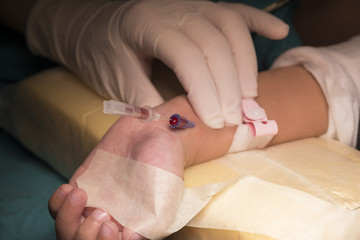

One of the most commonly used procedures in hospitals is cannulation. The process includes inserting a cannula (tube) into a duct, passage or cavity to deliver or withdraw fluid, other material, or blood sample. There are various kinds of cannulation in medical practice, including oral/nasal and IV (Intravenous).
IV cannulation is most frequently in use. In this, a cannula is inserted inside a vein for venous access. Access enables blood sampling, fluid/drug administration, parenteral nutrition, blood products, and chemotherapy. Here’s a simple guide to understanding the proper way of conducting IV cannulation.
How Does IV Cannula Work?
The IV cannulation procedure comprises four steps: explanation and collecting consent, preparation, process, and aftercare.
Explanation and consent
Before inserting the tube, the medical personnel must confirm patients’ identification, explain the purpose of the IV cannula procedure, describe the risks associated with it, review their medical history, and discuss the cannula location.
Preparation
Decontaminating your hands, wearing fitting non-sterile gloves, and an eye protection device is Cannulation 101. The gear prevents direct exposure to blood splashes. Place a tourniquet on the patient’s arm (preferably the non-dominant one), and select an insertion site.
Now, thoroughly cleanse the location with an alcohol swab for at least 30 seconds and then let it dry for another 30 seconds. Ensure you clean from the center of the site to the outwards and not touch once it’s sterile. A pillow under the patient’s arm can make the experience more comfortable.
Process
Aim for first-stick success, and insert the needle at an angle of roughly 30 degrees on the puncture location. Carefully observe the blood flashback and continue to insert an extra 1-2 millimeters to allow the catheter to enter the vein. The second flashback, blood between the needle and the catheter, confirms catheter’s entry into blood vessels.
Close the IV catheter’s lower end when you bring out the needle. To fulfill the purpose of I.V. cannulations in one go, ensuring the IV catheter’s proper placement is essential. Flush the catheter with some saline and check for any resistance or signs of extravasations around the site. Right after removing the needle, dispose of it in a sharps bin, discard the gloves and decontaminate your hands.
Aftercare
Inspect the patient for discomfort and the catheter for any fragmentation. After removing the catheter, put a gauze pad on the insertion site.
Types of I.V. Cannulas
I.V. cannulas uses can be broadly categorized into three sections: peripheral, central line, and midline. In peripheral IV cannulation, the forearm or hand’s peripheral veins are used for aspirating blood. Usually, patients in emergency rooms or surgeries receive this. For injecting antibiotics or long-term parenteral nutrition, central line IV is used. Patients requiring IV therapy for less than a month and more than five days receive a midline catheter.
Various applications of administering medications, IV fluids, or extracting samples, also depend on IV cannula sizes and uses. The following list mentions the IV cannula sizes, codes, and use cases.
- 14 G (Orange): The IV tube offers a flow rate of 240 ml/min. The cannula finds application in blood transfusion, rapid fluid replacement, and major surgeries.
- 16 G (Grey): The tube’s flow rate is 180 ml per minute. The 16 G and 14 G have similar functions.
- 17 G (White): With a blood flow proportion of 130 ml, the 17 G helps in rapid fluid replacement, surgical scenarios, and transfusion.
- 18 G (Green): Common functions for the IV tube include transfusion, trauma, and fluid replacement.
- 20 G (Pink): The tube with a flow rate of 60 ml helps in IV infusion.
- 22 G (Blue): First of the three IV cannula sizes for children, 22 G is perfect for pediatric blood infusion and transfusion and oncology and elderly patients.
- 24 G (Yellow): The tube is used for routine blood transfusion in neonates, children, and older patients.
- 26 G (Purple): With a flow rate of 13 ml, the cannula is best suited for pediatrics and neonates.
I.V. Cannula Sizes
| Colour Code | Orange | Grey | White | Green | Pink | Blue | Yellow | Violet | |
| Gauge | 14G | 16G | 17G | 18G | 20G | 22G | 24G | 26G | |
| Catheter Ext. (Diameter X Length MM) | 2.20 X 2.00 X45 | 1.70X45 | 1.50X 45 | 1.20X38 | 1.20X45 | 1.00X32 | 0.80X25 | 0.70X19 | 0.60X19 |
| Water Flow Rate M L/M IN | 310X290 | 200 | 140 | 105 | 100 | 64 | 38 | 16/22 | 12/15 |
Cannulation procedure requires professional training because if not done appropriately by considering the right size, specification, and puncture site, complications can arise.

